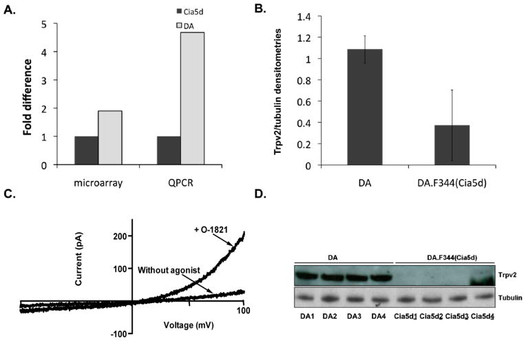
Figure 1. Trpv2 is functional and expressed in increased levels in invasive FLS from DA rats.
(A) Microarray analysis and QPCR confirmation showing increased mRNA levels of Trpv2 in DA FLS compared with DA.F344(Cia5d) (n=6 per group). (B) Trpv2 protein levels were also increased in DA FLS, compared with DA.F344(Cia5d), as shown on densitometries (normalized for tubulin±S.D) from Western blot (panel D). (C) FLS obtained from DA rats with PIA were patch-clamped in the absence of agonist, and after perfusion with O1821 30 μM, which induced channel opening. At positive potentials, the current is outward (upward), matching the Trvp2 pattern, thus confirming that FLS have functional Trpv2 channels. Currents are representative of 3–5 cells isolated from 3 different rats with PIA (n = 11 cells total). (D) Western blot image of four different DA and four different DA.F344(Cia5d) FLS cells lines (each from a different rat) demonstrating significantly higher Trpv2 protein levels in DA FLS (2.9-fold; densitometries on panel B).

