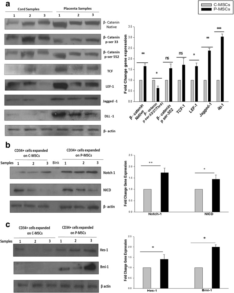
Fig. 7.

P-MSCs displayed higher Wnt activity leading to collateral increase into Notch signaling in the expanded CD34+cells. a Higher expression of native β-catenin. Phosphorylated β-catenin (ser-552) was found in the P-MSCs. Higher expression of phosphorylated β-catenin (ser-33/34Thr41) in C-MSCs. The expression of downstream targets of Wnt, TCF, LEF-1, jagged-1 and δ-like one (dll-1) was higher in P-MSCs than C-MSCs. The graph represents the fold change in the expression after normalization to β-actin. b Sort purified CD34+ cells from P-MSCs. Co-cultures showed significant up regulation of Notch I and intracellular domain of Notch (NICD). The quantitation of the same is represented as a graph. c The CD34+ cells expanded on the P-MSCs exhibited higher expression of Hes-1 and Bmi-1, downstream target of Notch, quantitatively represented as a graph. Data are represented as mean ± standard deviation from three different samples. *p ≤ 0.05, **p ≤ 0.01, and ***p ≤ 0.001. P-MSCs placenta-derived mesenchymal stem cells, C-MSCs cord-derived mesenchymal stem cells
