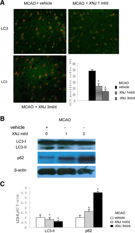
Fig. 6.

XNJ attenuated autophagy induced by cerebral ischemia. a The LC3-positive cells were analyzed using immunofluorescence 1 d after MCAO (upper panel, original magnification, 200×), and comparison of the percentage of LC3 positive cells among the experimental groups was exhibited (lower panel). Data are mean ± SEM of values obtained from 6 brains. b The levels of LC3 and p62 were analyzed by Western blot analysis. c Comparison of the density of LC3-II, p62/β-actin band among the experimental groups was showed. Data are the mean ± SEM of values obtained from 6 brains. *, p < 0.05, compared with the group of MCAO rats treated with vehicle
