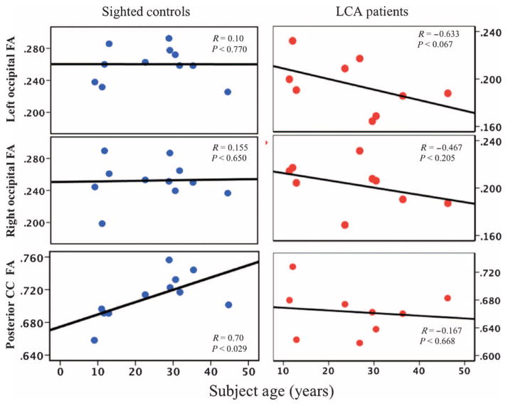
Fig. 2. Spearman correlations for occipital fractional anisotropy with age in LCA2 patients and sighted controls.
Shown are Spearman correlation analyses for white matter microstructural abnormalities within the right and left occipital cortex and posterior corpus callosum (CC) with age for LCA2 patients and sighted controls. LCA2 patients demonstrated similar correlations to those for sighted controls for the right occipital fractional anisotropy (FA), but the left occipital fractional anisotropy negatively correlated with age, thus demonstrating a continuous decline of the microstructural white matter in the left primary visual area for these patients. However, the posterior corpus callosum fractional anisotropy correlations with age were noticeably different for LCA2 patients compared to sighted controls. The absence of positive correlations for fractional anisotropy and patients’ age in the splenium of the corpus callosum for LCA2 patients may be due to the progressive nature of the disease, signifying the decline in communication between the two visual cortices over time and the reduction in the number of fibers crossing the splenium, which connects the left and right occipital cortices and enables binocular vision.

