Fig. 1.

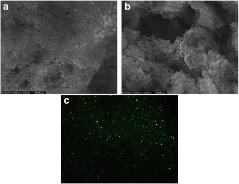
Scanning electron microscope image of free (a) and peptide loaded (b) PLGA nanoparticles (Magnification 5000x). Fluorescence microscope (40×) image of FITC + peptide loaded nanoparticles (c)

Scanning electron microscope image of free (a) and peptide loaded (b) PLGA nanoparticles (Magnification 5000x). Fluorescence microscope (40×) image of FITC + peptide loaded nanoparticles (c)