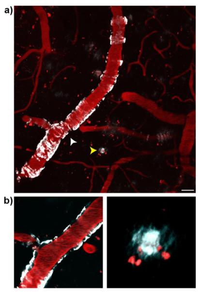
Fig.3.
Two-photon in vivo images of CRANAD-28 labeling in a 9-month-old APP/PS1 mouse. (a) Images were taken through a thinned-skull window 15min after i.v. infusion of the dye. Both CAA and amyloid plaques were labeled with CRANAD-28. (b) Lower panels show zoomed-in single focal plane examples of CAA (white arrowhead, left panel) and amyloid plaque (yellow arrowhead, right panel). Blood vessels were labeled with Texas-red dextran (70,000 MW). Red punctate signals are auto-fluorescence intracellular structures. Scale bar: 25 μm.

