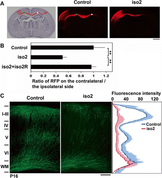
Fig. 5.

Role of mRbfox1-iso2 in the axon growth of cortical neurons in vivo. a pCAG-RFP was electroporated with control pSuper vector (control) or pSuper-mRbfox1-iso2 (iso2) into cerebral cortices at E14.5. Coronal sections were prepared at P3. Hematoxylin staining of a slice was also shown. Bar, 1 mm. b Quantitative analyses of the ratio of the intensity of RFP-positive axons in the area (white) of contralateral cortex to that in the area (green) of ipsilateral one in a. Rescue experiments were also done by cotransfection with pCAG-Myc-mRbfox1-iso2R (iso2R). Error bars indicate SD; control (n = 5), iso2 (n = 5), iso2 + iso2R (n = 4); **p < 0.01 by Tukey-Kramer LSD. c Representative images of the terminal arbors of axons expressing GFP with pSuper vector (control) or pSuper-mRbfox1-iso2 at P16. Note that axons of the deficient neurons reached contralateral hemisphere at the time point, whereas they did not extend efficiently into the cortical layer structure. Densitometric analyses of GFP fluorescence intensity were also carried out. Blue (control) and red (iso2) lines, average; shadow, SD (control, n = 6; iso2, n = 6). Bar, 200 μm
