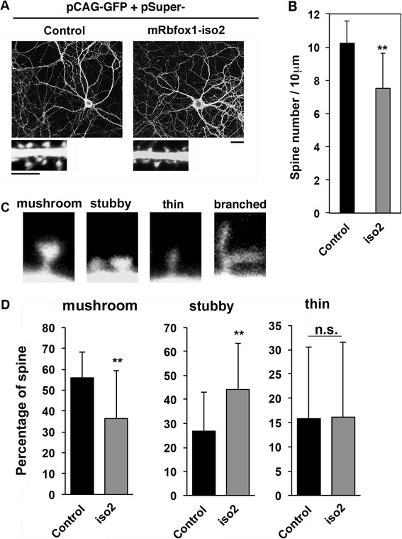
Fig. 7.

Role of mRbfox1-iso2 in the dendritic spine morphology of primary cultured hippocampal neurons. a Neurons were transfected with pβAct-EGFP together with pSuper vector (control) or pSuper-mRbfox1-iso2 (iso2) when isolated, fixed at 21 div and stained for GFP. Bar, 20 μm. Magnified images of dendrites are shown. Bars, 10 μm (upper panels) and 5 μm (lower panels). b Quantitative analyses of density of dendritic spines for each condition in a. Error bars show SD of the results from control (n = 17) and iso2-transfected neurons (n = 23). Experiments were repeated three times with similar results and representative data were shown. **p < 0.01 by Student’s t test. c Typical examples of mature (mushroom) and immature (stubby, thin filopodia, and branch) spines. d Relative abundance of the four different spine types in neurons transfected with pSuper vector (control) or pSuper-mRbfox1-iso2 (iso2) was analyzed. Relative percentages of spine types were indicated in graphs. Error bars show SD of the results from 150 spines (control, 17 cells; iso2, 23 cells). Experiments were repeated three times with similar results and representative data were shown. **p < 0.01 by Student’s t test. Note that branched spine was hardly detected under the conditions used (less than 2 % of total spine number)
