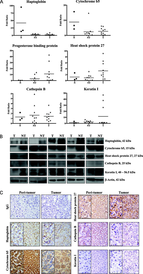
Fig. 4. Relative gene and protein expressions of the six protein markers in human liver tissues.
(A) Real time PCR was performed using total RNA isolated from normal human livers (N) (n = 3), non-tumor (NT) (n = 10) and tumor (T) (n = 10) liver tissues from HCC patients to examine the gene abundance. The relative gene expressions of haptoglobin, cytochrome b5, progesterone binding protein, heat shock protein 27, cathepsin B, and cytokeratin I were presented by fold ratio difference. The mean value of each category was also shown accordingly.
(B) Western blot was performed to reveal the protein levels of target proteins in peri-tumor (NT) and corresponding tumor (T) tissues of HCC patients. 10 pairs of NT and T clinical specimens were included and 5 representative sets of western blots are shown herein.
(C) Immunohistochemistry was performed to reveal the localizations and expressions of the target proteins in peri-tumor and corresponding tumor tissues of HCC patients. The primary antibodies targeting haptoglobin, cytochrome b5, heat shock protein 27, cathepsin B, cytokeratin I, or the respective IgG (negative control) were used at 1:100 dilution to reveal the corresponding immunoreactivities. A commercial antibody against progesterone membrane receptor component 1 is not available and thus not included. Representative micrographs of each antibody and selected IgG isotype control were shown. Magnification, ×200 (insert ×400).

