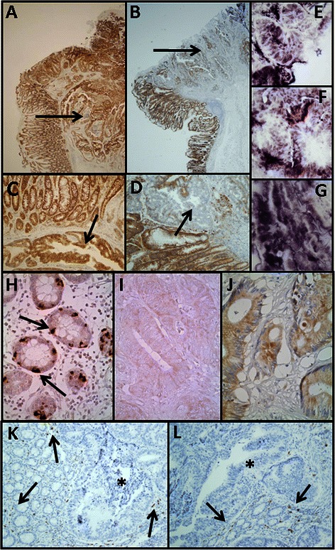
Fig. 2.

Immunolocalization of cell HSPGs. a-g Histological localization of syndecan-1 expression. a-d Localization of syndecan-1 in transition area between normal colon mucosa and tumor using immunohistochemistry. a, c Non-metastatic tumor; arrows indicate the retention of staining in the tumor region; b, d Metastatic tumor; arrows indicate the loss of staining in the tumor region, magnification A and B 20X, magnification C and D 100X; e-g CISH for syndecan-1 in CRCs. e Normal tissue; f Non-metastatic tumor; g Metastatic tumor; magnification 400X. h-j. Immunolocalization of glypican-1 expression. h, Normal mucosa. Intense staining appears in cells with neuroendocrine differentiation (arrows), whereas other epithelial cells show weak to moderate staining. i Non-metastatic CRC showing extremely weak staining. J Metastatic tumor showing weak staining levels; magnification 400X. k-l Immunolocalization of mast cells using CD117 antibody. Localization of mast cells in transition area between normal colon mucosa and tumor. k Non-metastatic CRC; l Metastatic CRC; magnification 200X. Arrows indicate the staining of mast cells in the normal mucosa. The asterisks show the tumor area, in which there is no detectable labeling
