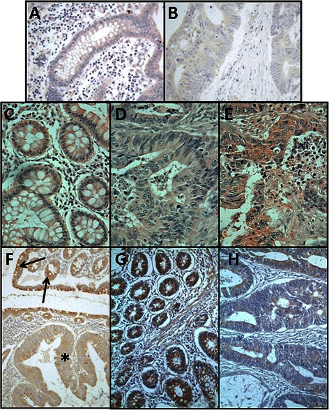
Fig. 5.

Immunolocalization of genes involved in the biosynthesis of HS and CS chains. a, b Immunolocalization of XYLT1 expression. a Normal mucosa showing weak to moderate staining. b Non-metastatic CRC, in which there is no detectable labeling; magnification 400X. c-e Immunolocalization of NDST1 expression. c Normal mucosa showing weak to moderate staining in the cell cytoplasm. d, Non-metastatic CRCs showing a decrease in the intesity of the staining. e Metastatic CRCs showing a similar staining intensity to that of healthy tissue. Magnification 400X f Localization of CHST12 expression in transition area between normal colon mucosa and tumor. Arrows indicate the staining of cells in the normal mucosa. The asterisks show the tumor area, in which the intensity of the labeling diminishes. Magnification 400X. g, h Immunostaining of UST. g Normal mucosa showing cytoplasmic staining in epithelial cells. h Non-metastatic CRC showing a notable decreasing in labeling. Magnification 400X
