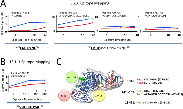
Fig 3. gH/gL epitope mapping by HDX-MS.
Curves of peptide deuterium uptake in the presence and absence of Fab are reported in red and blue, respectively. (A) Analysis of 3G16 binding identifies two peptides in the C-terminal region of gH presenting a deuterium incorporation reduction upon binding (peptides 677–684 and 705–725). The portion of the epitope present in the peptide 705–725 is narrowed to the sequence 705–708 since the two peptides 709–725 and 711–725 do not present any difference in deuterium uptake upon binding. (B) Analysis of 13H11 binding identifies a single peptide localized to the central portion of gH (peptide 238–247). (C) HCMV gH/gL complex model in ribbon representation showing the interaction sites with the neutralizing antibodies 13H11 (yellow circle) and 3G16 (orange circle), this study, and the previously identified MSL-109 binding site (green circle) [28].

