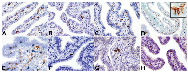
Figure 4. Cellular populations of the innate immune system in the normal fallopian tube.

Sections are from FFPE normal fallopian tube (A–H) and stained for CD11c (A), CD68 (B) and CD163 (C). D–E illustrate CD163(blue)/CD11c(brown) double immunostaining showing the presence respectively of CD11c+CD163− cells in the intraepithelial compartment (D; insert showing intraepithelial cytoplasmic projections of dendritic cells) and of CD11c+CD163+ cells admixed to CD11c−CD163+ cells in the lamina propria (E); F illustrates CD207/Langerin+ intraepithelial Langerhans cell; G illustrates occasional CD303/BDCA2+ plasmacytoid dendritic cells in vessels of the lamina propria; H illustrates intraepithelial CD56+ NK cells. Sections are counterstained with Meyer’s haematoxylin. Magnification: 40× (A–C and E–H) and 20× (D).
