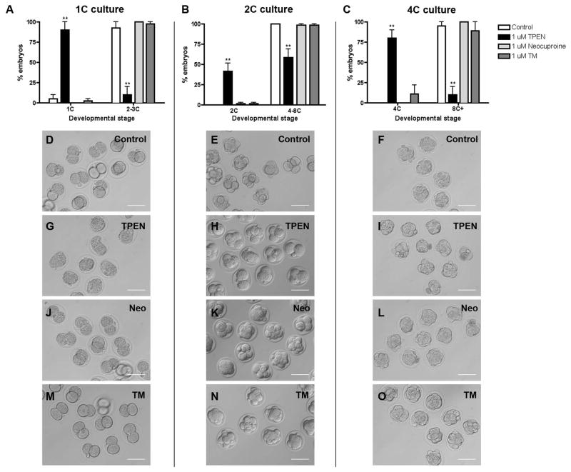
Figure 4. TPEN-induced arrest of preimplantation embryo development is zinc specific.
1C, 2C, and 4C embryos were cultured in media alone (control, D–F) or with 1 μM TPEN (G–I), 1 μM neocuproine (neo, J–K) or 1 μM tetrathiomolybdate (TM, M–O) for 24 h and developmental progression was scored. (A–C) The percentage of embryos at each developmental stage in each treatment condition was calculated. Experiments were performed in duplicate with 7–20 embryos per group. Results are reported as the mean ± SEM. Asterisks represent statistical significance as compared to control embryos at that particular developmental stage (**p<0.001). Representative transmitted light microscopy images of embryos in each treatment group are shown. Scale bars = 80 μm. 1C = one-cell, 2C = two-cell, 4C = four-cell, 8C+ = eight-cell, morula and blastocyst.

