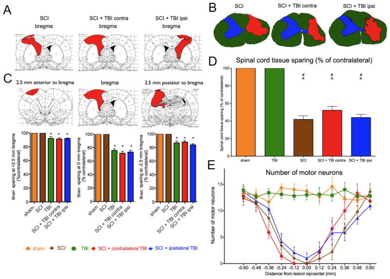
FIG. 5.
Both brain (A) and spinal cord (B) lesions were not significantly different in size or location between groups. The median lesions are shown for each group (A, B), and for each location.. (C). The median tissue sparing at 2.5 mm anterior to bregma, at bregma, and 2.5 mm posterior to bregma is shown below (*p<0.05 vs. Sham or SCI alone). (D) Spinal cord sparing at the epicenter of the lesion is shown (#p<0.05 vs. sham; *p<0.05 vs. TBI). (E) Motor neuron counts throughout the extent of the lesion show no significant differences between injury groups. SCI: spinal cord injury, TBI: traumatic brain injury.

