
Figure 1. Cell type-specific antigenic marker-based protocol for simultaneous tissue-specific isolation of PH, MΦ, and SEC from murine liver.
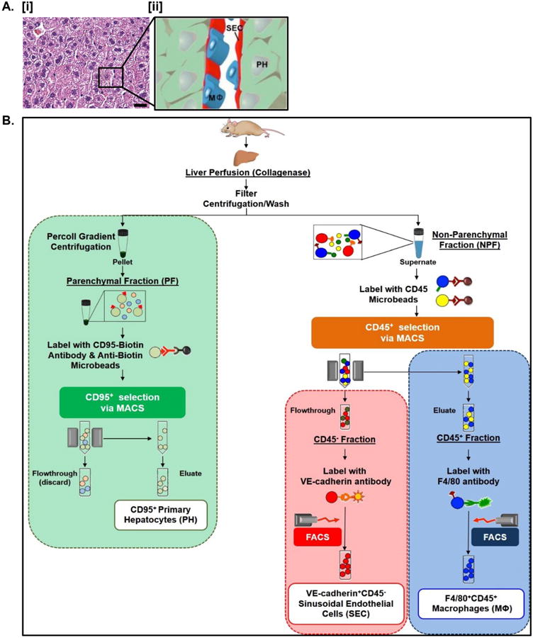
A. H&E stained paraffin-embedded sections of normal murine liver revealing normal endogenous tissue architecture in vivo [i]. The relationship between PH, MΦ, and SEC is highlighted in the accompanying schematic [ii]. Scale bars = 20 μm. See Table S3 for corresponding image magnification. B. Flowchart outlining experimental strategy for simultaneous isolation of PH, MΦ, and SEC.