
Figure 4. CD45- cells of the liver NPF are enriched for VE-cad+ SEC.
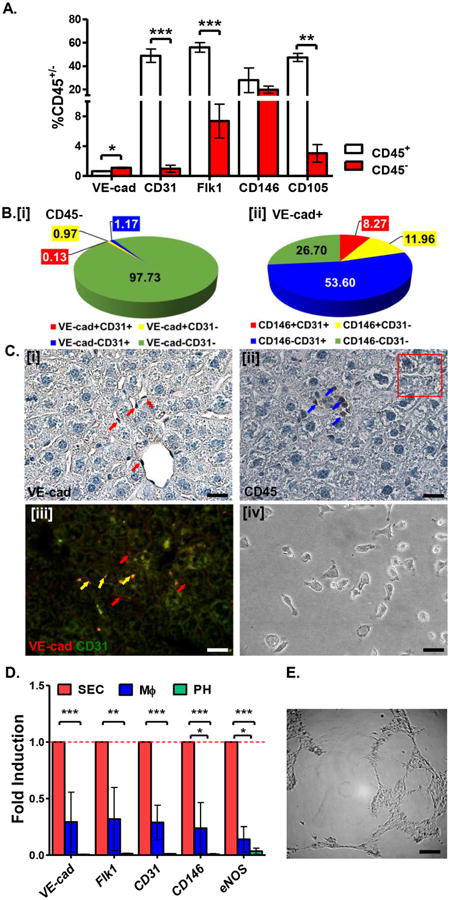
A. Percentage of liver NPF cells expressing SEC surface markers within the CD45+ and CD45- fractions. B. Distribution of SEC surface marker expression on CD45- cells from liver NPF. C. Immunostaining of murine liver sections demonstrates that cells lining the sinusoids express VE-cad [i] but not CD45 [ii]. VE-cad+ cells also co-express CD31 [iii]. Isolated tissue-specific VE-cad+CD45- cells demonstrate characteristic endothelial cell morphology in vitro [iv]. Scale bars = 20 μm. See Table S3 for corresponding image magnification. D. Relative expression (fold induction=1) of SEC-associated genes is enriched in VE-cad+CD45- cells (red bars) versus PH (green bars) and MΦ (blue bars) co-isolated from normal murine liver. E. VE-cad+CD45- SEC exhibits capillary tube-forming activity in vitro. Scale bar = 200 μm. See Table S3 for corresponding image magnification.