Figure 2.
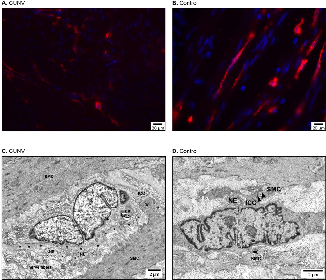
(A,B) Representative images showing depletion of the ICC network in the gastric smooth muscle in a CUNV patient (A) compared to control (B). The red signal is Kit immunoreactivity marking ICC; the blue signal is 4’,6’-diamidino-2-phenylindone counterstain marking cellular nuclei. (C,D) Electron microscopy images showing mild ultrastructural abnormalities in a CUNV patient (C) compared to control (D). Labeled structural components: thick basal lamina (small astericks), nerve endings (NE), smooth muscle cells (SMC), lamellar body (LB), ICC-SMC contact (arrows), and peg-and-socket junction (large astericks).

