Figure 5.
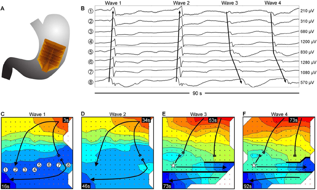
Abnormal slow wave initiation and conduction; isochronal intervals = 1.5 seconds. (A) Position of the array. (B) Representative electrograms from positions indicated in C; propagation sequences are labeled based on their corresponding waves 1–4, shown in panels C–F. (C) Isochronal activation map. Slow wave activity propagated onto the array from the greater-curvature, colliding with an unstable focal activity on the distal portion of the array. (D) The unstable focal activity was consistent over a second cycle. (E) A new stable ectopic pacemaker emerged in the distal portion of the array (represented by a star), initiating retrograde propagation that collided with the uncoupled antegrade wavefront. Circumferential propagation was out-of-phase with distal activity that was propagating in the opposite direction circumferentially, resulting in a complete functional conduction block (thick black line). (F) Propagation repeated as described in E, with stability of the ectopic pacemaker and distal block that remained consistent through the end of the recording period. A frequency increase occurred between the unstable focal activity of C&D (bradygastric, 2.0 ± 0.1 cycles/min) and the stable ectopic activity of E&F (normal frequency, 3.2 ± 0.2 cycles/min). See Supplementary Video 2 for animation.

