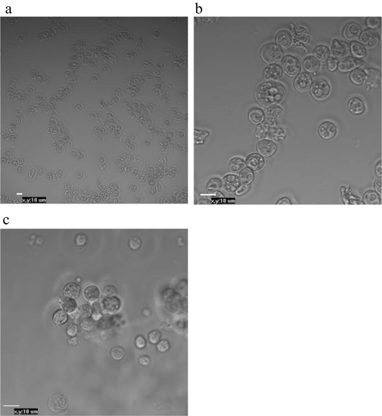
Fig. 2.
Morphological observations of PGCs that were isolated from the blood of embryos (stages 14–16 HH) and gonads of embryos (28–29 HH), purified and cultured for 24 h. bPGCs collected from between the 25 and 50 % Percoll dilution layers (a) (20× magnification). bPGCs after ACK buffer treatment (b). gPGCs are shown after trypsin digestion, Percoll purified and cultured for 24 h (c) (40× magnification)

