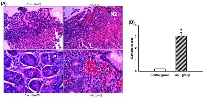
Fig 2. Histopathological examination of gastric mucosa sections.
A, Hematoxylin-eosin stained results showed appearance of the gastric mucosa in antrum region of the control group and HAPC-induced GML group (× 40 and × 400); B, The gastric mucosal damage score in control and HAPC-induced GML group. The mean scores were significantly higher in the HAPC-induced GML group when compared with a control group (P < 0.01, n = 3 per group).

