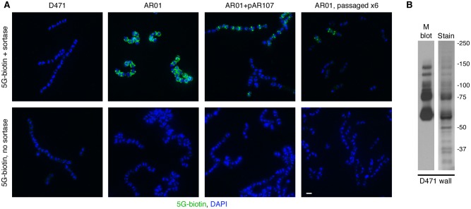
Fig 2. Missorted surface proteins accumulate in the sortase mutant AR01.
A. Wild type S. pyogenes D471, sortase mutant AR01, complemented AR01+pAR107, and a passaged AR01 variant, were diluted from overnight cultures 1:100 into fresh media (containing spectinomycin for AR01+pAR107), grown to log phase, fixed, attached to glass cover slides, and permeabilized with PlyC. The cells were blocked and incubated with pentaglycine-biotin (5G-biotin) in the presence or absence of purified S. aureus sortase A. The slides were washed, and pentaglycine-biotins molecules (attached to LPXTG motifs by sortase) were detected using FITC-streptavidin. DAPI was used to visualize DNA. The scale bar represents 1 μm. Note that expression of sortase from pAR107 is lower than the wild type, explaining the presence of intact LPXTG motifs. B. The cell wall fraction of log-phase D471 cells (solubilized with PlyC in PBS 30% raffinose) was separated by SDS-PAGE. Duplicate gels were stained with GelCode blue or processed for Western blot using the 10B6 monoclonal antibody. The most prominent bands on the stained gel correspond to M protein.

