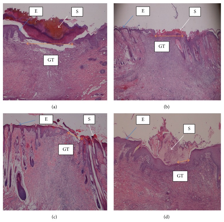
Figure 2.
Histology of wound tissue at day 14 after wounding in rats, stained with hematoxylin and eosin and dressed with (a) 0.2 mL of vehicle, gum acacia, wide wound area (orang arrow); (b) 0.2 mL of Antrodia camphorata (100 mg/mL) moderate wound area compared to vehicle; (c) 0.2 mL of Antrodia camphorata (200 mg/mL), smaller wound area compared to vehicle (orange arrow); (d) 0.2 mL of Intrasite gel, very small wound area compared to vehicle (orange arrow). S = scab, E = epidermis, and GT = granulation tissue (magnification 20x).

