Figure 1.
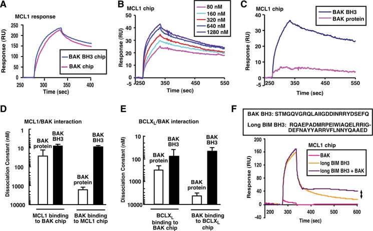
MCL1 and BCLXL preferentially bind activated BAK. (A) SPR of 300 nM MCL1ΔTM binding to immobilized BAKΔTM or BAK BH3 peptide. (B) SPR (relative units) of different concentrations of BAK BH3 peptide binding to immobilized MCL1ΔTM. (C) Direct comparison of 320 nM BAKΔTM and 320 nM BAK BH3 peptide binding to immobilized MCL1ΔTM. (D,E) Based on the SPR assays, the affinities of MCL1ΔTM (D) and BCLXLΔTM (E) for BAKΔTM and BAK BH3 peptide were calculated. Error bars indicate ±sd of three independent experiments using different chips and different protein batches. (F) SPR of MCL1ΔTM binding to 500 nM BAKΔTM, 500 nM BIM BH3, or 500 nM BAKΔTM + 500 nM BIM BH3. The double arrow indicates increased resonance due to BAK activation. The box at the top of F indicates peptides used in this figure.

