
Figure 6.
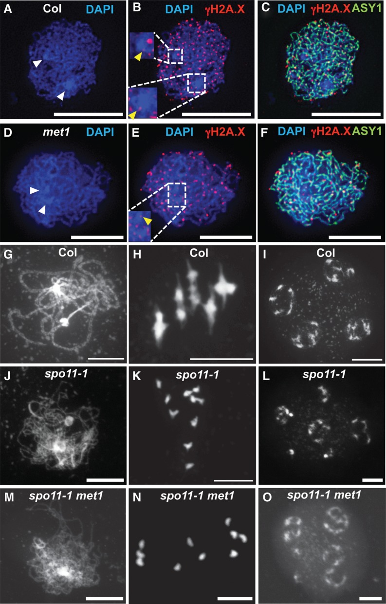
Meiotic DNA DSB foci are unchanged in met1. (A–F) Localization of ASY1 (green) and γ-H2A.X (red) in wild-type (A–C) and met1-3 (D–F) nuclei at leptotene. DNA was stained with DAPI (blue). Densely DAPI-staining chromocenters are marked with white arrowheads. The inset boxes show magnifications of the centromeric regions, with yellow arrowheads marking γ-H2A.X foci localized within the DAPI-dense regions (Supplemental Fig. 2). Bars, 10 μm. (G–O) DAPI-stained meiotic chromosome spreads of wild-type (G–I), spo11-1 (J–L), and met1 spo11-1 (M–O) nuclei at late prophase I (G,J,M), metaphase I (H,K,N), and tetrad (I,L,O) stage. Bars, 10 μm.