
Figure 7.
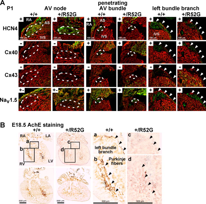
Expression of positive- and negative AV nodal, penetrating AV bundle and left bundle branch markers in P1 hearts, and AchE activities in E18.5 developing hearts. (A) Representative images of serial tissue sections positively (+) or negatively (−) stained with HCN4, connexin 40, connexin 43, and Nav1.5 in AV node, penetrating AV bundle (traced by white dots) and left bundle branch (arrowheads) from P1 mice. (B) Representative images demonstrating AchE activity (brown) in left bundle branch and ventricular trabeculations (Purkinje fibers) from serial tissue sectioning of control Nkx2-5+/+ and Nkx2-5+/R52G (+/R52G) hearts, is weaker in Nkx2-5+/R52G hearts. Areas a–d selected on the left are shown enlarged on the right. RA, right atrium; RV, right ventricle; LA, left atrium; LV, left ventricle; AS, atrial septum; IVS, interventricular septum.