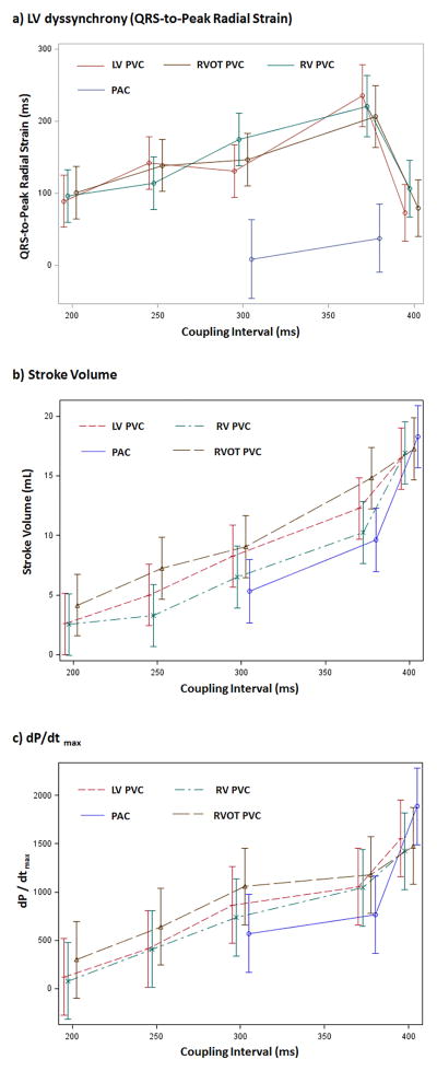
Figure 1.
(a) LV dyssynchrony (QRS-to-Peak LV radial strain), (b) stroke volume, and (c) dP/dtmax in premature ventricular beats (PVC) by different coupling intervals (200, 250,300, 375ms) and rapid ventricular paced beats (400ms) from RV, RVOT and LV free wall, as well as premature atrial beats (PACs) at 200ms (R-R interval 290–430ms). RV= RV apex, RVOT= RV outflow tract, LV = LV free wall.

