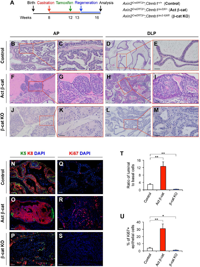
Figure 5. β-catenin is required for castration-resistant Axin2-expressing cells to expand luminal lineages during prostatic regeneration.
(A): A scheme of the experimental designs for analyzing castration-resistant Wnt/β-catenin-responsive cells in Axin2CreERT2/+;Ctnnb1+/+ (control), Axin2CreERT2/+;Ctnnb1(ex3)fl/+ (Activated β-cat), and Axin2CreERT2/+;Ctnnb1(ex2-6)fl/fl (β-cat KO) mice. (B–M): H&E staining of AP and DLP tissues isolated from different mice. (N–P): Co-immunofluorescence staining of K5 (green), K8 (red), and DAPI, and Ki67 and DAPI in different mouse models. Representative images of AP were shown. (T): Ratios of luminal (K8+) versus basal (K5+) cells in prostate tissues isolated from mice received testosterone treatment for 3 weeks. (U): Percentages of Ki67+ epithelial cells in the above prostate tissues. Error bars indicate standard deviation. *, P < 0.05; **, P < 0.01 by Student’s t tests. Scale bars, 50 µm.

