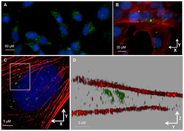
Fig 1. Liver fluke granulin internalized by H69 cholangiocytes.
(A) Widefield (deconvolved) micrographs showing the lateral (xy) overview of live H69 cholangiocytes imaged after 18 h incubation with Alexa Fluor 488-conjugated rOv-GRN-1 (green) and Hoescht nuclear stain (blue). (B) With further magnification of fixed cells the labeled rOv-GRN-1 was evident among the cytoskeletal actin network (red) of numerous cells with DAPI (blue) stained nuclei. (C) 3D-SIM lateral (xy) overview image of a well-separated individual cholangiocyte stained as in panel B. (D) Rendered axial (yz) view of boxed inset in (C) showing rOv-GRN-1 (green) present between the apical and basal actin filaments (red) of the cholangiocyte (DAPI channel omitted). Additional material shown in S1 Fig and S1 Movie.

