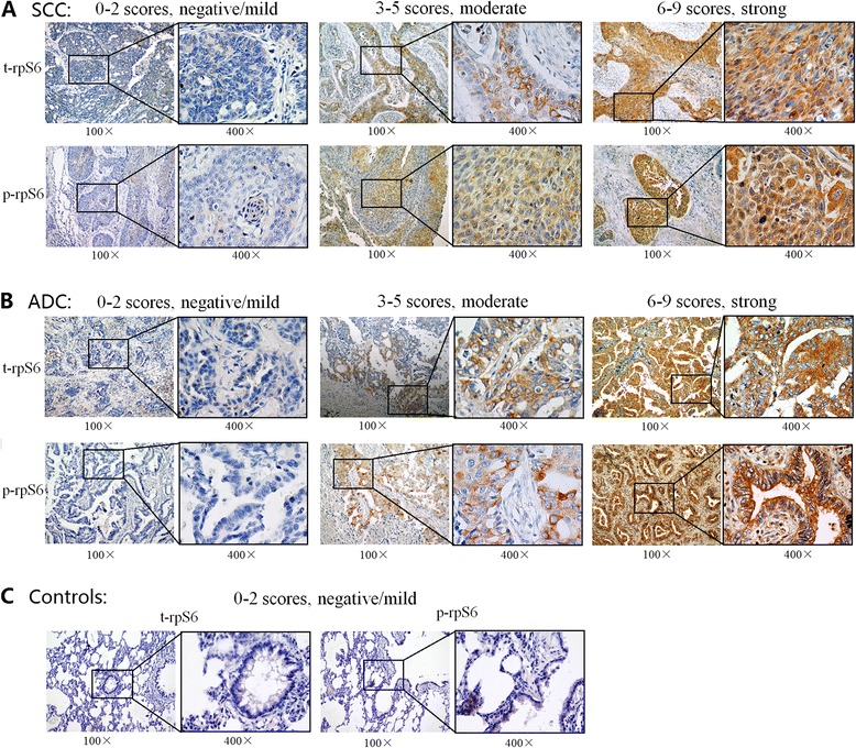
Fig. 1.

Expression of t-rpS6 and p-rpS6 were both highly expressed in NSCLC tissues. Representative t-rpS6 and p-rpS6 immunohistochemical stainings in squamous cell carcinoma squamous cell carcinoma (a) and adenocarcinoma (b) tissues showed their abundant accumulations in the cytoplasm of tumor cells, which were divided into negative/mild, moderate and strong grade. However, their expressions in non-tumor lung tissues (c) were negative or mild. Each low magnification (100 ×) were paired with a high magnification (400 ×) for clear observation. t-rpS6: total rpS6; p-rpS6: phosphorylation of rpS6; ADC, adenocarcinoma; SCC, squamous cell carcinoma
