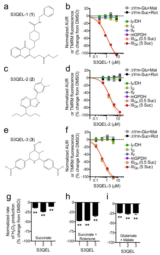
Figure 1. Chemical screening using isolated mitochondria identifies suppressors of site IIIQo superoxide production.
(a – f) Structures of S3QELs 1-3 and dose-response curves against two ΔΨm and six H2O2 endpoint screening assays (n = 1). Mean IC50 values against site IIIQo superoxide production were 0.75, 1.7, and 0.35 μM for S3QELs 1-3, respectively. (g – i) Effects of S3QELs 1-3 on the steady state rate of H2O2 production measured using the Amplex UltraRed assay (normalized mean ± SE, n = 3 biological replicates). **p < 0.01 versus DMSO in each condition; one-way ANOVA with Dunnett’s posttest. Glu, glutamate; Mal, malate; Suc, succinate; Rot, rotenone; IF/DH, site IF plus NADH-linked matrix dehydrogenases.

