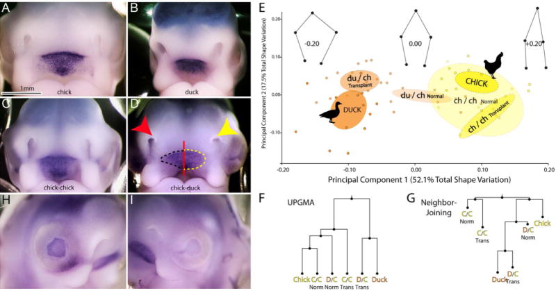
Figure 3. Effect of brain transplantation on morphology and Shh expression at stage 22.

(A) Frontal view of a chick and (B) duck embryo at HH 22 after in situ hybridization to illustrate Shh expression at this stage. (C) In a chick-chick chimera no obvious difference in Shh expression is observed on the control (right side of embryo) and chimeric (left side of the embryo) sides of the embryo. (D) In a duck-chick chimera the nasal pit (yellow arrow) on the chimeric side (left side of embryo) is rounded and appears more duck like at this stage. On the control side (right side of embryo) the nasal pit (red arrow) appears as a slit, which is similar to the chick. Similarly, Shh expression on the operated side resembles the rounded duck-like expression pattern (outlined in yellow), while on the control side, the expression domain remains host-like (outlined in black). (E) Principal Components Analysis (PCA) of nasal pit morphology demonstrates that chicken (yellow, n=7 embryos, n=14 sides) and duck (orange, n=7, embryos, n=14 sides) separate along PC1 (51.5% total shape variation). Chimeric side nasal pits of experimental embryos (du/ch transplant n=15 embryos, n=30 sides) overlap ducks, while the normal side of experimental embryos (du/ch normal) tends to be more chicken-like, matching predictions and qualitative observations. Control animals comprised of chick-chick transplants (ch/ch transplant) overlap with normal chicks (Chick). Wireframes show landmark displacements along PC1. Ellipses show 95% confidence intervals of group means. (F) Both UPGMA and (G) Neighbor-joining trees of Procrustes distances between group means links Duck-Chimera to the exclusion of a Chicken-Normal. In F and G, the distances between all groups were significantly different (p<0.05) except for normal chickens and chick-chick chimeras (p>0.05). (H) On the control side of the embryo eye development is more advanced as evidenced by the dark pigmentation of the pigmented retina compared to (I) the lighter pigmentation in the pigmented retina on the chimeric side of the embryo.
Scale bar=1mm.
