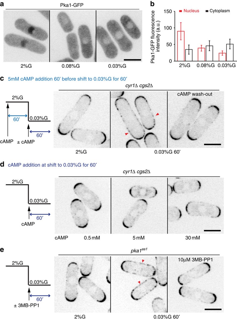
Figure 2. Pka1 is active in low glucose to promote Pom1 side-localization.
(a) Maximum intensity spinning disk projection of Pka1-GFP in wild-type cells grown in 2% or 0.08% or 0.03% glucose for 1 h. (b) Measurement of cytoplasmic and nuclear Pka1-GFP levels in cells as in a (n>20). Experiments were performed thrice and quantification of one is shown. (c) Medial spinning disk confocal section of Pom1-tdTomato in cyr1Δcgs2Δ cells incubated with 5 mM cAMP in 2% glucose for 1 h (left) and shifted to 0.03% glucose for 1 h with the same amount of cAMP (middle) or without cAMP (right). Arrowheads indicate Pom1 at cell sides. (d) Medial spinning disk confocal section of Pom1-tdTomato in cyr1Δcgs2Δ cells grown in 2% glucose and incubated with increasing amounts of cAMP at the time of shift to 0.03% glucose for 1 h. (e) Medial spinning disk confocal section of Pom1-tdTomato in pka1-as1 cells grown in 2% glucose (left) and shifted to 0.03% glucose without (middle) or with 10 μM 3MB-PP1 (right). Arrowheads indicate Pom1 at cell sides. Scale bars are 5 μm. Error bars are s.d. For c–e, representative images from two independent experiments are shown.

