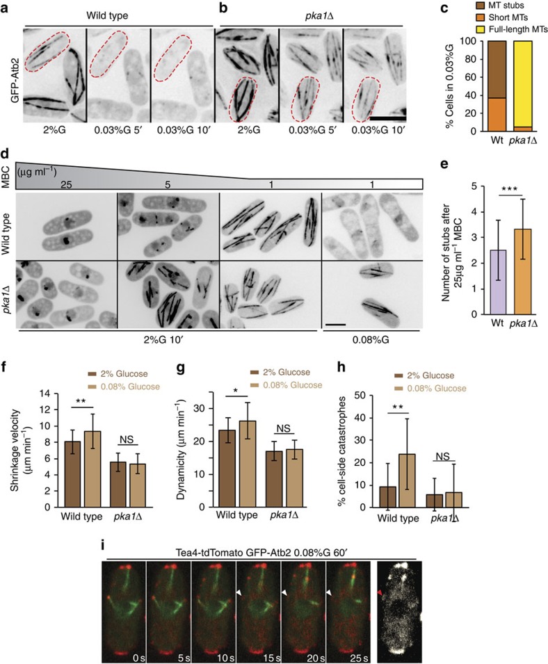
Figure 4. Pka1 negatively regulates microtubule stability.
(a) Epifluorescence deconvolved maximum intensity projection images of wild-type cells expressing GFP-Atb2 grown in microfluidic chambers in 2% glucose and 5 and 10 min after shift to 0.03% glucose. (b) GFP-Atb2 in pka1Δ cells as in a (2% glucose and 0.03% glucose after 10 min). Representative images from three independent experiments are shown. (c) Percentage of wild-type (n=68) and pka1Δ (n=59) cells with indicated microtubule organization in 0.03% glucose for 10 min in microfluidic chambers. (d) Maximum projection of spinning disk confocal images of wild-type and pka1Δ cells expressing GFP-Atb2 treated with the indicated concentrations of MBC for 10 min in 2% or 0.08% glucose. Representative images from two independent experiments are shown. (e) A graph showing the number of microtubule stubs left after 10 min 25 μg ml−1 MBC treatment (n>45 cells). P=0.001. (f) Mean microtubule shrinkage velocity in wild-type and pka1Δ cells grown with 2% glucose or 0.08% glucose for 1 h (n>27 microtubules). (P=0.008; P=0.56). (g) Mean microtubule dynamicity in wild-type and pka1Δ cells grown with 2% glucose or 0.08% glucose for 1 h (n>27 microtubules; P=0.031; P=0.54). (h) Percentage of microtubule catastrophes occurring at the cell sides in wild-type and pka1Δ cells (n>100 catastrophe events in >16 cells) grown as in e. (P=0.002, P=0.926). (i) Time-lapse imaging of Tea4-tdTomato and GFP-Atb2 in wild-type cells shifted to 0.08% glucose for 1 h acquired on the spinning disk confocal microscope. The first six images are maximum projections of two Z-sections. The last image is a projection of the three time points shown after microtubule catastrophe. Arrowheads indicate Tea4 presence at the lateral cell cortex after microtubule catastrophe. Representative images from four independent experiments are shown. Scale bars represent 5 μm. Error bars are s.d. Statistical significance was derived using student's t-test.

