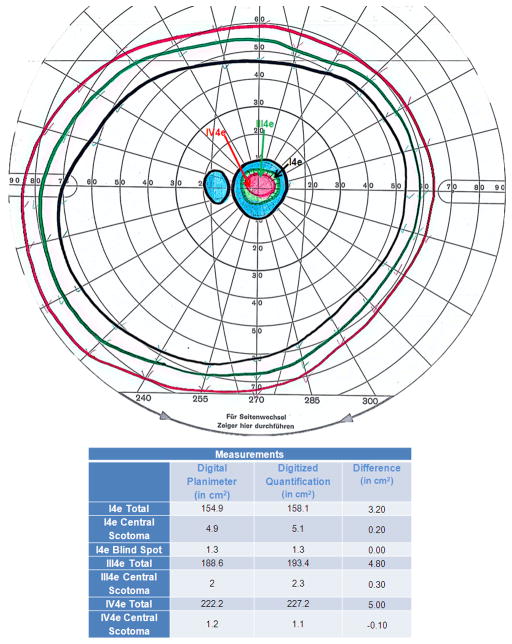
Figure 2. Comparison of Traditional versus Novel Digital GVF Quantification.
Quantification of the total extent of peripheral fields, physiologic blind spot, and central scotoma were performed by a single observer in square centimeters (cm2) for the I4e, III4e, and IV4e isopters using digital planimetry and Adobe Photoshop for a 35-year-old female patient with Stargardt disease. The absolute differences between the two methodologies are shown in the table.

