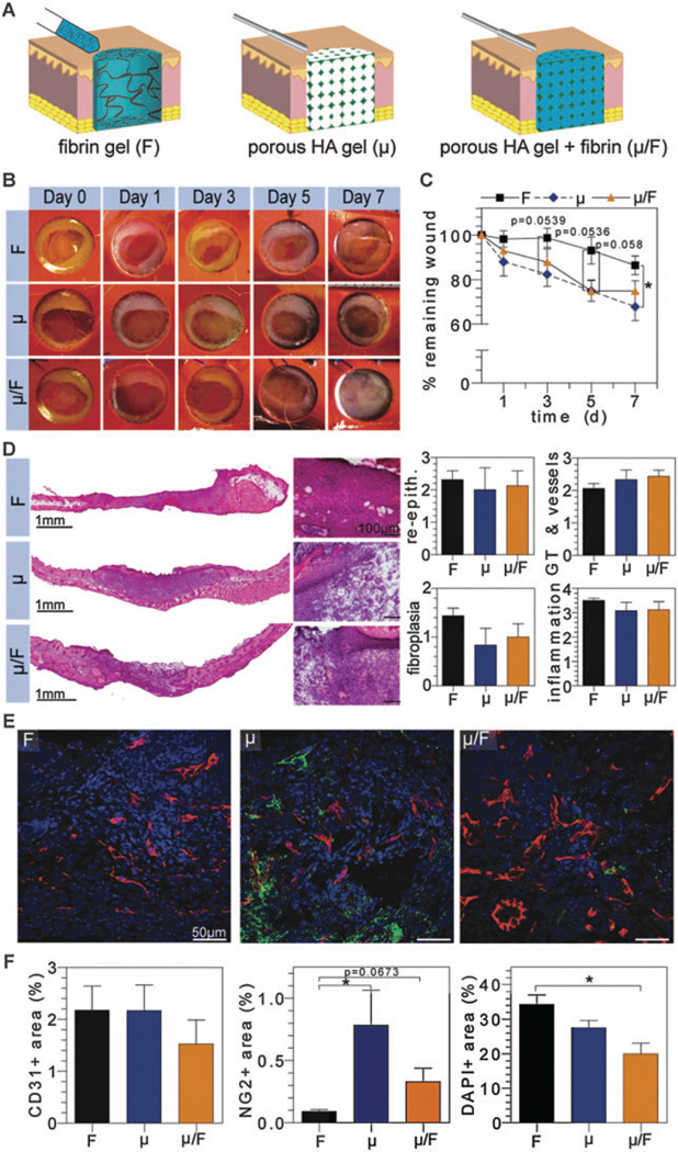
Fig. 2.
Evaluation of composite natural scaffolds in vivo. (A) Schematic representation of wound treated with fibrin (F), microporous HA (μ), or composite (µ/F) hydrogels. Digital images of wounds were taken at regular time intervals for wound closure analysis (B and C). H&E stained wounds depict granulation tissue formation and were scored by a pathologist for re-epithelialization, granulation tissue/vessel formation, fibroplasias, and inflammation (D). OCT embedded sections were stained for vascular cell populations (E), where CD31+, NG2+, and nuclei imaged in red, green, and blue, respectively, and quantified via imageJ (F).

