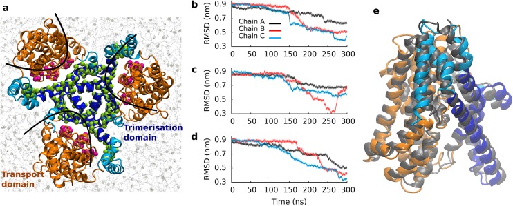
Fig 1. Global structure of GltPh.
(a) Membrane embedded trimeric transporter structure, seen from the extracellular site. The transport domain is shown in orange, the trimerization domain in light (membrane exposed) and dark blue (without membrane contacts). The same color code is applied to all figures. The backbone atoms used as pulling groups are highlighted in green and purple. (b-d) Time evolution of transporter geometry is shown of three independent simulations, measuring the structural deviation of each protomer individually (chain A in black, chain B in red, chain C in blue) from the inward-facing crystal structure. (e) Overlay of the inward-facing crystal structure in gray (PDB ID: 3KBC) and the closest conformation extracted from the SMD simulation. The RMSD between the structures was 0.35 nm.

