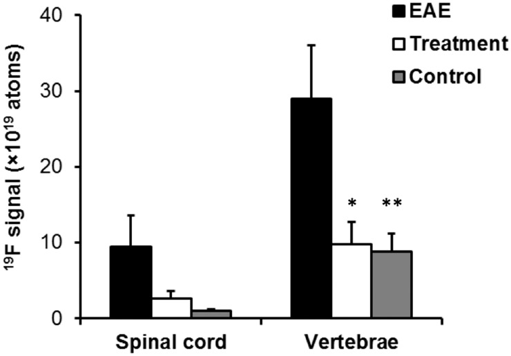
Fig 2. In vivo quantification of inflammation load in the spinal cord and vertebral bone marrow in EAE rats.
19F signal in the spinal cord and vertebral bone marrow is significantly increased in EAE rats compared to wild-type rats (black vs. grey bars; p < 0.05 indicated by *). 19F signal in the spinal cord and vertebral bone marrow of EAE rats is significantly decreased by prophylactic treatment with cyclophosphamide (black vs. white bars; p < 0.05 indicated by **). The apparent number of fluorine atoms was measured by integrating the signal in ROIs and normalizing the results to the signal in a reference capillary containing a known concentration of fluorine, as discussed in Methods section.

