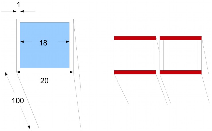
Fig 9. Geometry of the vacuole.
Left: a cell represented for computational purposes as a brick shape with a square cross section. The vacuole is in blue. Measurements in microns. Right: we consider lateral coupling in the horizontal direction. The cytoplasmic layers in red can be regarded as the routes for lateral flux.

