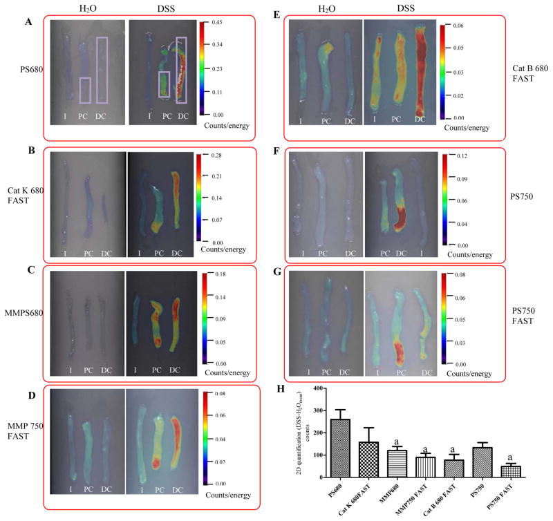
Figure 1. Strong and significant activation of all probes were detected in distal colon of DSS treated mice by ex vivo FRI imaging.
Representative 2D FRI images of fresh intestine from DSS-treated mice versus water controls are shown in Figure 1A through Figure 1G. Rectangle ROIs (in purple color) were placed on distal colon and distal half of proximal colon to quantify fluorescence signal (counts). (H) 2D Quantification of fluorescence signals in colon from DSS-treated animals (n=4–9 animal/group). Data are mean±SEM of total counts per colon ROI in each DSS treated animal injected with each of the indicated probes after subtraction of the mean value in water controls.
I:ileum; PC: Proximal colon; DC: Distal colon
a: p<0.05 vs. PS 680 (ProSense 680)

