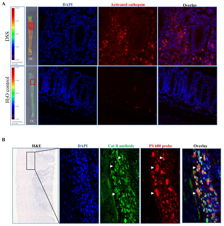
Figure 2. ProSense 680 probe activation co-localized with cathepsin B antibody immunofluorescence staining.
(A) Cathepsin enzyme activation was detected only in colon from DSS treated animal, but not in colon from water control animal. (B) Co-localization of ProSense 680 probe activation and cathpsin B antibody by immunostaining. White arrows indicate the overlap of enzyme activation and cathepsin B antibody staining.
DC: Distal colon

