. Author manuscript; available in PMC: 2015 Oct 24.
Published in final edited form as: Neurol Res. 2012 Jun 28;34(7):677–684. doi: 10.1179/1743132812Y.0000000064

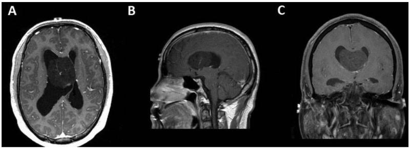
Figure 1. A 45-year-old male presenting with headache and fatigue with a 4.6 cm transverse × 4.8 cm anteroposterior × 3.8 cm craniocaudal, heterogeneously-enhancing mass in the anterior body of the right lateral ventricle. Patient had prominent lateral ventricles and received an intra-operative ventricular catheter. (A) Minimally enhancing lesion on an axial T1-weighted post-contrast image. (B) Minimally enhancing lesion on a coronal T1-weighted post-contrast image. (C) Minimally enhancing lesion on a sagittal T1-weighted post-contrast image.

Figure 1