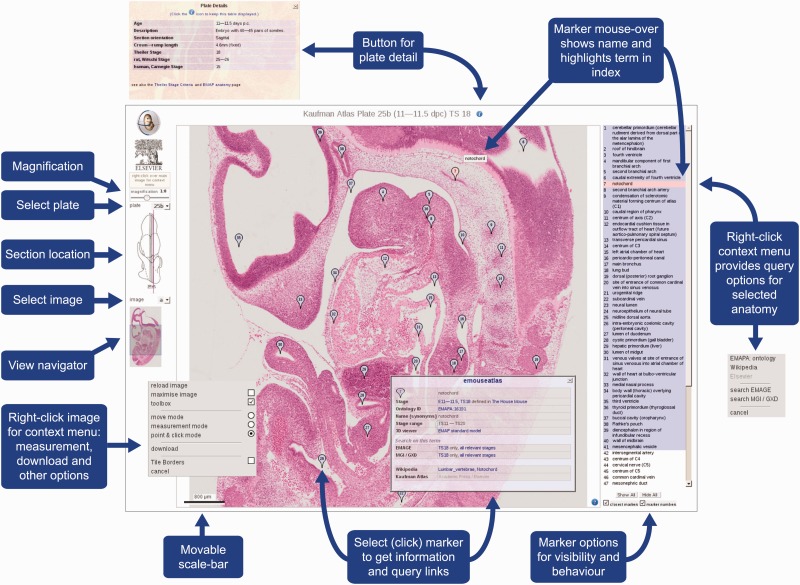
Figure 1.
eHistology Atlas IIP3D Viewer Details. This figure shows the various options and navigation tools available within the IIP3D viewer used in the eHistology Atlas. Plate 25b (image a) is shown, with the colour section image displayed in the central panel at 1:8 magnification, the anatomy labels are listed on the right, and navigation tools shown on the left. Clicking on a numbered flag opens up the floating pop-up window containing internal and external links. Image is provided with permission from eMouseAtlas.

