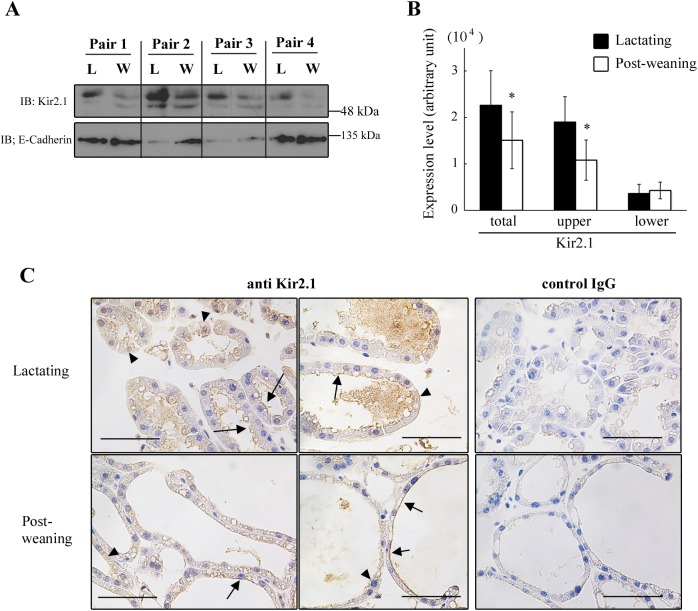
Fig 5. The effects of forced weaning on the protein expression of Kir2.1 in MS cells.
A, B: expression of Kir2.1 protein in the microsomal fraction of MS cells. Dams of a similar age, nursing duration, and litter size were paired. The cell isolation and enrichment of microsomal fraction were performed simultaneously for each pair. The lysate (35 μg of protein) of the microsomal fraction from lactating (L) and post-weaning (W) MS cells were separated with SDS-PAGE and immunoblotted with the anti-Kir2.1 and anti-E-cadherin antibody (A). Two bands around 50 kDa are shown. The densities of the bands were measured (B). Data represent mean ± SE (n = 4 for each). *, p < 0.05 vs. lactating, using paired t-test. C: localization of Kir2.1 in mammary glands. Sections of lactating (upper panels) and post-weaning (lower panels) mammary glands were immunostained with anti-Kir2.1 antibody and control IgG (for negative control). Arrows indicate cells stained at the apical membrane. Arrowheads indicate cells with diffuse staining in the cytoplasmic region and at the basolateral side of the cells. Black bars show 50 μm.

