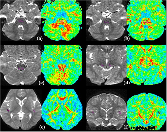
Fig 1. Representative DT imaging of the ROIs.

(a) the trapezoid body, (b) superior olivary nucleus, (c) inferior colliculus, (d) medial geniculate body, (e) the auditory radiation, (f) the white matter of Heschl's gyrus, (square box) the selected ROI.
