Fig. 8.

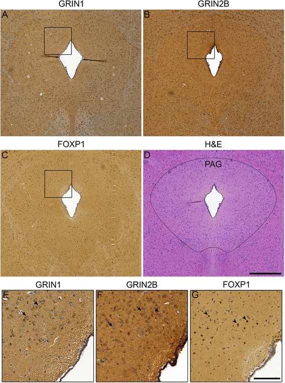
Midnight blue hub genes are expressed in the PAG of the bat brain. Protein expression pattern of (a) GRIN1 (b) GRIN2B and (c) FOXP1 in the bat PAG. d Haematoxylin and Eosin (H&E) staining of the bat PAG to show tissue structures - the boundary of the PAG has been demarcated with a dotted line. Higher magnification photos show (e) GRIN1 and (f) GRIN2B in the cytoplasm of cells within the PAG as indicated by the dark brown staining (arrow), whereas (g) FOXP1 could be found in nuclei (arrow head) as expected for a transcription factor. A regulatory relationship for GRIN2B and FOXP1 has previously been shown in the mouse. The overlapping expression pattern in regions of the bat PAG, suggest that in a subset of cells FOXP1 may be able to regulate GRIN2B expression. The scale bar represents 500 μm for a-d (d) and 125 μm for e-g (g)
