
FIGURE 1.
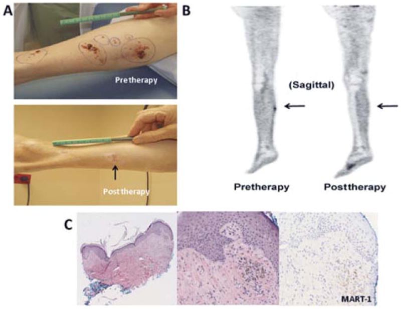
An example of a patient who was CR by FDG-PET/CT with persistent superficial skin lesions on physical examination ultimately found to be pigment only on histology; the patient was classified as a CR. A, Photographs demonstrate the persistence of a pigmented lesion (arrow) 12 weeks post-ILI. B, Sagittal FDG-PET/CT images pre-ILI (left) showing increased FDG uptake within the right calf and at 12 weeks post-ILI (right) showing resolution of the hypermetabolic soft tissue nodule within the right calf. C, Punch biopsy of the pigmented lesion at 12 weeks post-ILI. Left, a low-power magnification shows pigmented cells in the dermis (4X), the middle and right photographs taken in high power (20X) highlight the MART-1 negative pigmented cells supporting the interpretation of melanin-laden macrophages.