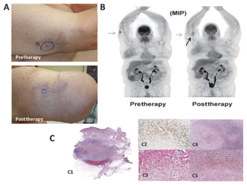
FIGURE 3.

An example of a patient who had palpable but decreased in size nodules on physical examination and persistent FDG activity ultimately determined to be fibrotic nodules in-filtrated with pigment-laden macrophages and no tumor; this patient was classified as a CR. A, Photographs demonstrate absence of lesions at 12 weeks post-ILI but a persistent palpable nodule. B, FDG-PET/CT maximum intensity projection images pre-ILI (left) showing hypermetabolic superficial soft tissue nodules along the right forearm and at 12 weeks post-ILI (right) showing a presence but decrease in number and metabolic activity of right upper extremity nodules (an example is highlighted by the gray arrows). The black arrow points to fat necrosis that can be seen on FDG-PET/CT after treatment. C, Wide local excision of the palpable lesion at 12 weeks post-ILI. On the left (C1), a scan of the whole section mount to demonstrate the location of the nodule, at right there is a medium (C2, 2X) and high (C3, 10X) power view of the pigmented cells. Immunohistochemical stains performed in this specimen show that the cells are negative for MART-1 (C4, 10X) and positive for CD163 (C5, 10X), supporting the interpretation of melanin-laden macrophages.
