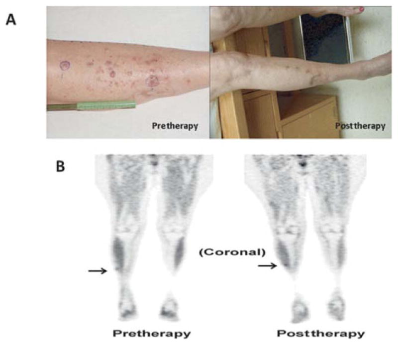
FIGURE 4.

An example of a patient who had a CR by physical examination but had persistence of FDG activity on PET/CT determined by histology to be melanoma; this patient is classified as a partial responder. A, Photographs demonstrate resolution of lesions at 12 weeks post-ILI. B, Coronal FDG-PET/CT images pre-ILI (left) showing mild focal FDG uptake in the right anterior leg (arrow) and at 12 weeks post-ILI (right) showing unchanged low-level FDG activity in the right anterolateral shin. Biopsy of this FDG spot using needle localization was consistent with melanoma.
