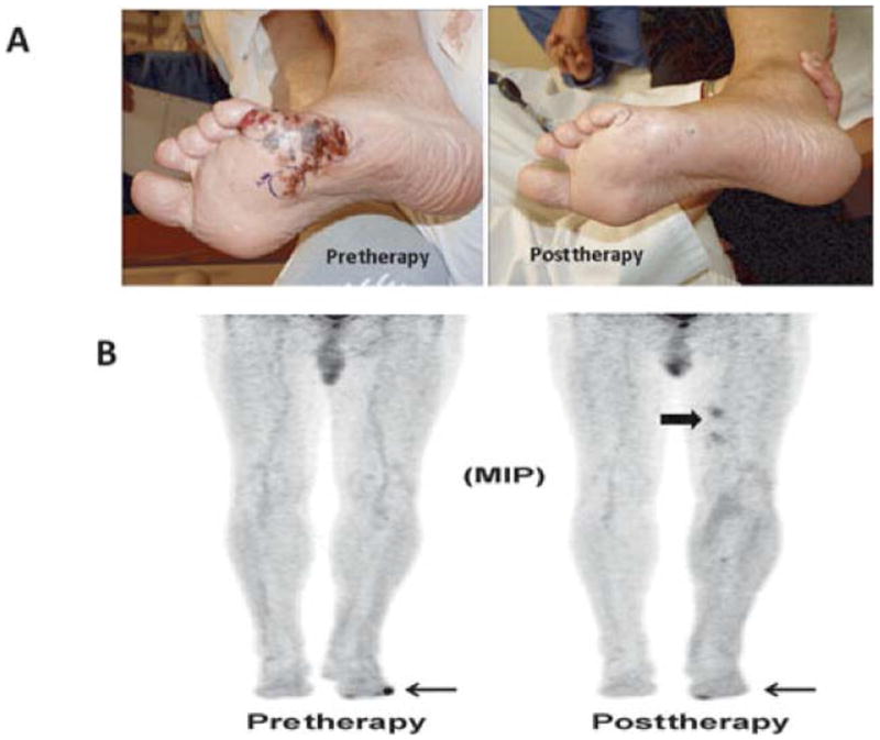
FIGURE 5.

An example of a patient who had a partial response by physical examination but complete resolution of FDG activity on PET/CT; this patient was classified as a partial responder. A, Photographs demonstrate a partial response of lesions at 12 weeks post-ILI. Biopsy of this clinical evident lesion was consistent with melanoma. B, Maximum intensity projection FDG-PET/CT images pre-ILI (left) showing a hypermetabolic focus in the lateral aspect of the left foot (arrow) and at 12 weeks post-ILI (right) showing interval resolution of the hyper-metabolic focus of activity along the lateral aspect of the left foot. Note there is some activity on the medial portion of the foot near the great toe, which was unrelated to the melanoma. Also noted is myonecrosis not infrequently seen after ILI in the upper thigh (block arrow), which is considered a false-positive FDG-PET/CT finding.
